








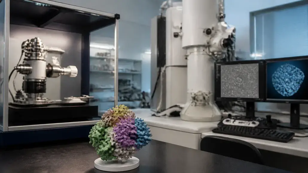
X-ray crystallography usually fits best when you need fast, high-resolution structures of a protein that crystallizes, especially with many bound ligands, while cryo-EM fits best for large complexes, membrane proteins, and flexible assemblies that resist crystallization. Both methods are now central to structural biology. Of the 20,974 structures deposited to the Protein Data Bank in 2025, 53.1% came from X-ray crystallography and 45.3% from 3D electron microscopy, according to the RCSB PDB Q1 2026 deposition report.
That near-even split makes the choice harder for research teams. Picking the wrong method can cost months of construct work, sample optimization, and instrument time before anyone realizes the project is stuck. Budgets and timelines rarely allow for a second attempt from scratch.
The good news is that a handful of practical questions about your target and your goals usually point clearly toward one method, or toward a smart combination of both.
How X-Ray Crystallography and Cryo-EM Each Build a 3D Structure
X-Ray Crystallography: Reading Diffraction From Ordered Crystals
Crystallography starts with growing a well-ordered crystal of the protein. When an X-ray beam hits the crystal, millions of identical, aligned molecules scatter it into a diffraction pattern, which is then converted into an electron density map. Once a reliable crystal system exists, new ligand-bound structures can be solved in days, often at resolutions better than 2 Å.
The hard part is getting there. Construct design, protein purification, and crystallization screening can take weeks to months, and some proteins never crystallize at all. Teams without crystallization robotics or regular synchrotron access often turn to protein crystallography services for this stage, from construct optimization through to data collection and refinement.
Cryo-EM: Averaging Images of Frozen Single Particles
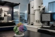
Single-particle cryo-EM skips crystals entirely. The sample is flash-frozen in a thin layer of vitreous ice, and an electron microscope records hundreds of thousands of images of individual particles in random orientations. Software sorts and averages these images into a 3D reconstruction.
Because particles are imaged one by one, cryo-EM can separate different conformational states from a single dataset. That makes it uniquely powerful for dynamic machines such as transporters, ion channels, and multi-protein complexes.
Cryo-EM vs X-Ray Crystallography at a Glance
| Factor | X-ray crystallography | Cryo-EM |
| Sample requirement | Well-diffracting crystals | Pure, homogeneous protein in solution |
| Ideal target size | Any size, including small domains | Generally above about 50-100 kDa |
| Typical resolution for drug targets | Often 1.5-2.5 Å | Often 2.5-3.5 Å, sometimes better |
| Membrane proteins | Possible, often difficult | A major strength |
| Multiple conformations | Usually one state per crystal form | Several states from one dataset |
| Ligand series throughput | High once soaking works | Improving, still lower |
| Main bottleneck | Crystallization | Grid preparation and microscope time |
Five Questions That Settle X-Ray Crystallography vs Cryo-EM
How Large and How Flexible Is Your Target?
Size is often the first filter. Small, soluble proteins and isolated domains below roughly 50 kDa give cryo-EM too little signal to align particles accurately, so crystallography is usually the practical route. Large complexes, viruses, and membrane assemblies point the other way, especially when flexible regions would prevent tight crystal packing.
Does Your Protein Crystallize, or Could It?
A previously published crystal form is a major head start. If related structures exist in the PDB, a similar construct and condition often work for your protein too. When initial screening does not result in well-diffracting crystals, performing a feasibility Cryo-EM study can be faster than continuing to optimize.
What Resolution Does Your Question Really Need?
Not every project demands the same detail. Match the method to the level of detail your decision depends on:
- Overall architecture and subunit arrangement: 4-6 Å is often enough, which cryo-EM delivers readily
- Side-chain positions and binding pocket shape: around 3 Å or better
- Precise ligand pose, water molecules and hydrogen-bond geometry: ideally below 2.5 Å, where crystallography remains the most consistent
How Many Ligand Structures Will You Need?
Structure-based drug design runs on volume. A medicinal chemistry program may need dozens or hundreds of protein-ligand complexes over its lifetime.
Crystal soaking systems can deliver these at a pace that matches weekly design cycles, and fragment screening campaigns routinely process hundreds of crystals. Cryo-EM ligand throughput is rising quickly, though each structure still typically demands more microscope time and processing effort.
Do You Need to See Several Conformational States?
Many proteins work by changing shape: transporters open and close, receptors switch between active and inactive states. Cryo-EM can capture several of these states from one sample, while a crystal usually locks the protein into a single conformation. For mechanism-focused research, that difference can decide the choice on its own.
In practice, teams often work through these questions in order:
- Check the PDB and literature for existing structures of the target or close homologs.
- Assess size, flexibility, and membrane association.
- Define the resolution and ligand throughput the project requires.
- Run a short feasibility phase with the most promising method.
- Keep the second method in reserve if feasibility stalls.
Combining Both Methods, With AI Models as the Starting Point
The sharpest research programs rarely treat the choice as permanent. A common pattern uses cryo-EM to solve the full complex and reveal how its parts fit together, then crystallography of a key domain to generate high-resolution ligand structures for chemistry. Each method covers the other’s blind spots.
AI structure prediction now sits upstream of both. Predicted models help in several practical ways:
- Designing constructs that trim disordered regions likely to block crystallization
- Solving crystal structures through molecular replacement when no experimental model exists
- Docking predicted models into lower-resolution cryo-EM maps to speed up interpretation
- Prioritizing which conformations or complexes are worth the experimental effort
Predicted models still miss details that matter for drug design, such as ligand-induced pocket changes and ordered water networks. Experimental structures remain the reference point for those decisions.
Matching the Method to the Decision Your Project Must Make
For most small-molecule drug discovery programs, ligand throughput decides the method, and that still favors X-ray crystallography whenever the target crystallizes. Cryo-EM earns its place where crystals fail, where the biology lives in large or membrane-bound assemblies, and where several conformations matter to the question being asked.
The two techniques are converging in capability year after year. Teams that define their resolution needs, ligand volume, and target behavior up front can choose with confidence, and keep the other method ready as a powerful complement.
Frequently Asked Questions
Is cryo-EM replacing X-ray crystallography?
No. Cryo-EM depositions are growing fast and now approach crystallography in annual numbers, but crystallography remains the main source of high-resolution protein-ligand structures. The two methods increasingly work side by side.
Which method should I choose for a small protein under 50 kDa?
X-ray crystallography is usually the better choice. Small proteins give cryo-EM too little signal for reliable particle alignment, although fusion partners, antibody fragments, or scaffolds can sometimes make cryo-EM possible.
Can I use cryo-EM for structure-based drug design?
Yes, and it is increasingly common for targets such as GPCRs, ion channels, and transporters. Plan for lower ligand throughput and check that the local resolution around the binding site is good enough to place the ligand confidently.
How long will it take me to get a structure with each method?
With an established crystal system, a new ligand complex can take days. A brand-new target may need weeks to months for either method, depending on how quickly you can produce stable protein, crystals, or good cryo-EM grids.
Which method is more expensive for my lab?
Building in-house cryo-EM capacity is far more expensive, since high-end microscopes and detectors require major investment and specialist staff. Per structure, costs depend heavily on how difficult the sample is, so many labs outsource either method rather than buying equipment.