













A scan can meet a site’s routine clinical standards and still leave a trial team with an unusable assessment. Across a global imaging network, that gap creates expenses that don’t sit neatly within the imaging budget: repeat visits, reconciliation work, and unresolved questions approaching database lock.
Phase III CNS Protocol Optimization brings those operational exposures into focus before they become recovery projects. In CNS clinical trials, imaging must support comparisons across sites and over time, not simply produce interpretable pictures. When study endpoints depend on those comparisons, acquisition differences become a concern for operational leadership as well as imaging specialists.
The planning question is therefore broader than the price of each scan. It concerns who owns consistency across a multi-year study, particularly when scanners, software, and site personnel change.
What Scanner Variance Really Costs a Phase III CNS Trial
The financial exposure lies beyond the original scan charge. Repeat visits, query cycles, missing timepoints, and submission-review questions represent different consequences, and they require different controls. Paying for another scan can’t recreate every lost assessment.
Standardization means specifying comparable outputs across scanner configurations, not distributing the same document worldwide. One global acquisition framework needs scanner-specific implementation, controlled exceptions, and a central owner responsible for compliance throughout the study. Qualification, change control, timely quality checks, and documented interpretation belong in the budget before first patient in.
Where Site-to-Site Imaging Variance Drains the Budget
Imaging variance rarely appears as one budget line. Its costs spread across site payments, data management hours, and schedule changes, making the original acquisition problem difficult to track financially.
Software Versions and Hardware That Don’t Match
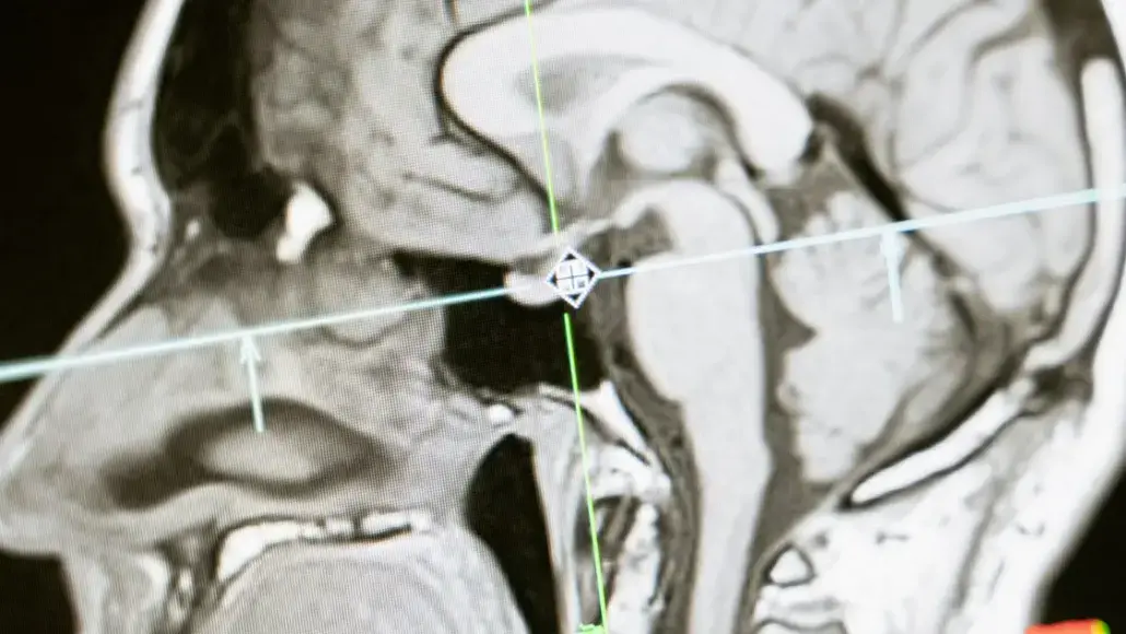
Scanner specifications deserve explicit treatment in trial design. For central nervous system (CNS) imaging, a network spanning different field strengths, coil configurations, and software releases shouldn’t assume that identical sequence names produce equivalent images.
The operational question is whether each configuration delivers images suitable for the planned assessment. Qualification therefore needs scanner-specific acquisition settings, rather than a written instruction that every site interprets independently.
Mid-study changes create a separate exposure. A software upgrade or hardware replacement requires documented assessment before subsequent scans enter the same longitudinal series. Otherwise, teams face retrospective queries about when the change occurred, which participants were affected, and whether their assessments remain comparable.
Those investigations consume resources already committed to patient recruitment and trial delivery.
Calibration Drift and the Re-Scan Cycle
Gradual scanner changes aren’t necessarily obvious during routine site review. Comparing quantitative measures across sites and timepoints through neuroimaging data analytics provides a way to investigate signal or geometric deviations that local checks haven’t identified. Without scheduled comparisons, affected scans could accumulate for weeks before investigation begins.
The financial consequence depends on when the problem surfaces. Detection during the assessment window leaves room to investigate and, where appropriate, repeat acquisition. Later detection leaves fewer recovery options.
A repeat scan creates another scheduling task, patient visit, and potential site charge. If the assessment window has closed, another appointment doesn’t automatically replace the missing endpoint observation. Budgets should distinguish recoverable acquisition failures from missing timepoints that additional spending can’t restore.
Why CNS Endpoints Magnify Small Acquisition Differences
For imaging assessments of brain metastases, lesion visibility and measurement deserve separate scrutiny. Acquisition requirements must support both the identification of eligible lesions and consistent measurement at subsequent visits.
RECIST 1.1 and RANO-BM Need Comparable Scans
RECIST 1.1 and RANO-BM provide defined approaches to response assessment, but they aren’t interchangeable acquisition manuals. RANO-BM addresses brain metastases specifically and connects measurable-lesion assessment to contrast-enhanced imaging and acquisition resolution.
An operationally sound protocol doesn’t reduce either framework to a minimum lesion diameter. It specifies the applicable criteria version, slice-thickness requirements, measurement rules, and handling of lesions that aren’t measurable. Exact thresholds belong in the controlled imaging documents, checked against the published criteria before site activation.
The distinction matters because a change in lesion visibility shouldn’t be mistaken for a biological change. For CNS metastases trials using progression-free survival, acquisition-related uncertainty needs documented adjudication rather than an unexplained endpoint assignment.
Leptomeningeal disease also needs explicit assessment rules; a framework centered on discrete target-lesion measurements isn’t sufficient by itself.
When Imaging Data Weakens an NDA or EMA Filing
The submission concern is traceability: can reviewers reconstruct how images became endpoint results? For a new drug application (NDA) to the FDA or a marketing authorisation application to the European Medicines Agency, unexplained acquisition changes leave that question harder to answer.
The FDA’s Clinical Trial Imaging Endpoint Process Standards guidance provides a relevant framework for documenting imaging processes. Where FDA Oncology Center of Excellence guidance informs an oncology program, the protocol should identify the applicable recommendations rather than treat all CNS assessments alike.
Scanner variance doesn’t automatically determine regulatory approval. The defensible concern is whether unresolved differences undermine the reliability or reviewability of the evidence, prompting questions that require additional analysis and explanation.
How Standardized Protocols Keep Imaging Submission-Ready
 As the site-level risks illustrate, qualification should establish that each scanner can deliver the required acquisition before participant imaging begins. This provides the starting point for the ongoing checks described below.
Site Qualification and Phantom-Based Calibration
Test submissions demonstrate protocol execution, while phantom-based checks provide a controlled reference for evaluating specified scanner characteristics. These checks serve different purposes: a technically acceptable phantom result doesn’t establish that staff have followed every participant-imaging instruction.
Repeat qualification should follow a predefined schedule and relevant equipment changes. The change-control record needs to identify the affected scanner, implementation date, assessment performed, and authorization to resume study acquisition. That record limits retrospective investigation and helps prevent unresolved equipment changes from delaying database lock.
Centralized Quality Control and Independent Review
Central quality control should assess each incoming examination against the imaging requirements within a defined turnaround. The deadline needs to reflect the remaining visit window, including time for site contact, investigation, and any permitted repeat acquisition.
The workflow should distinguish three outcomes:
- An acceptable examination that can proceed to assessment.
- A correctable submission problem, such as missing files.
- An acquisition failure requiring documented clinical and operational review.
This distinction avoids treating every query as a reason for another patient visit.
Blinded independent central review then addresses interpretation, applying the specified response criteria consistently. It doesn’t repair deficient acquisition. Separating acquisition checks from endpoint interpretation creates a clearer audit trail for FDA review and internal reconciliation.
Leaner Protocols That Protect Recruitment and Timelines
Every required sequence should have a defined purpose tied to study endpoints, safety assessment, or necessary quality control. A sequence without such a purpose adds work without a clear evidentiary return.
Removing unnecessary acquisition requirements is a trial design decision, not a site-level shortcut. The revised protocol must still preserve everything needed for the planned assessment.
For CNS clinical trials, this discipline also keeps patient recruitment discussions grounded in the actual visit burden. Sites need a feasible acquisition schedule, realistic transfer deadlines, and clear escalation rules. A shorter protocol isn’t automatically better, but an essential-only protocol avoids paying for data the study won’t use.
Treating Imaging Consistency as a Budget Line, Not a Fix
Phase III CNS Protocol Optimization therefore treats consistency as a planned operating expense rather than an emergency recovery task. That approach doesn’t promise zero rejected scans or guaranteed regulatory acceptance.
It establishes a more defensible position: acquisition requirements are explicit, deviations have accountable owners, and the evidence linking images to endpoint results remains traceable. The decision is to fund that structure early rather than rely on retrospective repair.